Dental CT scan in Barcelona: CBCT on a Carestream CS 8200
Corona Dental takes dental CT scans in its own imaging room at Entença 69, on a Carestream CS 8200 3D Advance Edition. The exposure itself takes seconds, the dentist opens the 3D reconstruction on screen during the same appointment, and you leave with the file. No separate trip to an imaging centre, no waiting until tomorrow for a disc. We work in English, Spanish, Russian and Ukrainian.
<1 minin the scanner
€80–150depending on the area covered
Same dayread on screen, file in hand
DICOMopens in any dentist's software
What a dental CT scan costs
| Item |
Price |
| Dental CT scan (CBCT) in our imaging room |
€80–150 |
| Periapical X-rays |
included in the appointment |
The range reflects how much is being covered. One or two teeth need a small field of view, and that is the lower end. Both arches in full, which is what implant and orthodontic planning calls for, need a wide field, and that is the upper end. Which one your case needs is decided before you go into the room, and the exact figure is quoted beforehand. Dental care provided for diagnosis or treatment is exempt from Spanish VAT, so the figure quoted is what you pay. Having it done commits you to nothing further: the file is handed over once it is paid for.
What dental CT scans cost around Barcelona
Worth knowing before you book anywhere, because the same examination is sold at quite different prices depending on who takes it.
| Where |
Typical range |
What that buys |
| Private health booking platforms |
€69–160 |
Low headline price, but nobody interprets the images and the referral has to be sorted separately |
| Dental clinics with their own CBCT |
€80–150 |
Imaging and interpretation in one place, in one appointment |
| Hospitals and imaging centres |
€140–170 |
A formal written radiology report, aimed at complex cases and medical referrals |
Market guidance for Barcelona in 2026, set out so you can compare. These are not our fees — ours are in the table above. Everything else is on the price list.
CBCT or dental CT scan: which term is right
Most people search for a dental CT scan, which is why this page is headed that way. The machine standing in a dental clinic, though, is a CBCT — cone beam computed tomography. In Spain you will also see it advertised as TAC dental, TC de haz cónico or escáner 3D; in English, cone beam CT, 3D dental scan or simply CBCT scan. They all mean one thing.
The distinction is not pedantry. A hospital CT sweeps a narrow fan of X-rays across the body while the couch moves through the gantry, stacking slices as it goes. A CBCT fires a cone-shaped beam at a flat panel detector and makes a single rotation around your head; the computer builds the whole volume from that one pass. That is where the three things patients notice come from — seconds rather than minutes, a far lower dose, and sitting up instead of lying down — and it is also why the machine fits inside a dental clinic rather than a hospital radiology department.
What comes out is not a flat picture but a block of data the dentist moves through slice by slice, in any plane: axial, coronal, sagittal, and curved along the line of the arch. On an ordinary X-ray everything is superimposed and one root can hide the one behind it. In three dimensions each is looked at separately and measured in real millimetres.
A CBCT scan is not the hospital CT scan
This deserves its own section, because in Spain both tests are called TAC and someone searching for a dental CT scan often lands on an imaging centre's website and starts preparing for the wrong thing entirely.
| Point |
Dental CBCT |
Hospital CT |
| Area covered |
Jaws and the structures around them, nothing else |
Any region of the body |
| Beam shape |
Cone beam, one rotation, flat panel detector |
Fan beam, successive slices as the couch advances |
| Position |
Sitting or standing, chin on a rest |
Lying on a couch that moves into the gantry |
| Time |
Seconds |
Ten minutes upwards |
| Contrast |
Never used |
Often injected |
| Dose |
Much lower |
Higher, varies with the protocol |
| Preparation |
None |
Sometimes fasting; bloods first if contrast is used |
| Where |
In the dental clinic |
Hospital or imaging centre |
| Who reads it |
The dentist who will treat you |
A radiologist, who issues a written report |
If imaging has been asked for to place implants, plan orthodontics or take out a wisdom tooth, what is meant is a dental CBCT. None of those jobs need a hospital scanner and there is no reason to pay the difference. A medical CT belongs to other situations: tumours, extensive sinus disease, facial trauma with multiple fractures, anything spreading beyond the jaws.
Do you need a referral?
Yes — and it is worth saying plainly, because almost no clinic website mentions it. Spanish radiation protection law, Royal Decree 601/2019 on justifying and optimising the use of ionising radiation, says no X-ray examination may be carried out unless a professional entitled to request it has justified it first. The principle will be familiar to anyone who has had imaging done in the UK: it is the same justification duty that sits at the heart of IR(ME)R. In practice it means a dental CT scan is not something you can simply buy over the counter. A dentist, surgeon or doctor has to indicate it in writing.
That leaves two routes, and both work here.
1
You already have a referralBring it, in any language. We scan the field your dentist has asked for and hand you the file so they can open it. You do not have to be treated here or move practice.
2
You have no referralThe indication is established at an appointment. A first appointment costs €50 and includes examining your mouth; if the case warrants a 3D study, it is done the same day, with no second trip.
Platforms selling imaging on its own meet the same requirement by adding a consultation, online or in person, to issue the paperwork. It gets paid for either way — just split across two invoices. Worth bearing in mind when you compare an advertised price with the final one.
That rule has a clinical consequence as well. Whoever justifies the exposure has to record what clinical question it answers, so nobody here covers both arches on the off-chance. Beforehand the dentist says what they expect to see and picks the smallest field of view that answers it. A smaller field means a smaller dose.
What the 3D images show
| Structure |
What appears |
| Available bone |
Height and width of the ridge in millimetres, plus bone density. Without it there is no deciding the diameter and length of an implant, or whether a graft is needed first |
| Maxillary sinus |
Where the sinus floor sits, how thick the lining is, what previous surgery left behind. This is what settles whether a sinus lift is required |
| Inferior dental nerve |
The exact course of the ID nerve canal through the lower jaw. This is the difference between a predictable wisdom tooth extraction and one carrying a risk of nerve injury |
| Roots and canals |
How many canals there really are, how they curve, which ones a previous root canal treatment missed |
| Infection at the root tip |
Granulomas and cysts, including those an ordinary X-ray cannot separate from the root in front of them |
| Fractures and resorption |
Vertical root fractures and internal or external resorption, both awkward to spot in two dimensions |
| Impacted teeth |
Buried wisdom teeth and canines, how they are angled, and how close they sit to neighbouring roots and to the nerve |
| Airway and jaw joint |
The upper airway and the shape of the condyles — information orthodontics and grinding cases need |
When a 3D image is needed
ImplantsMeasuring bone, choosing the implant, planning guided surgery
Wisdom teethSeeing how the roots relate to the nerve before going in
Root canal retreatmentFinding missed canals and infection that will not settle
OrthodonticsBuried teeth, skeletal anchorage, root resorption
Pain with no visible causeWhen an ordinary X-ray does not explain the symptom
Jaw surgeryPlanning on the real bone rather than an estimate
TraumaCracks and displacement that a periapical X-ray misses
What happens on the day
1
Jewellery offEarrings, piercings, hair clips, glasses, necklaces and any removable denture. Metal throws streaks across the image wherever it sits.
2
Getting into positionYou sit or stand. Your chin rests on a support and two side guides steady your head — they do not grip, they just stop you drifting.
3
The rotationThe arm makes one full turn around your head. Stay still and breathe normally; there is nothing to feel and nothing to hear beyond the motor.
4
ReconstructionThe computer assembles the images in under a minute. No film, nothing to develop.
5
Looking at it togetherThe dentist moves through the slices with you sitting there, points out what has turned up and explains what it changes about the plan.
For anyone without mobility difficulties, the whole thing — walking into the room, sitting down to look at the images — takes a few minutes.
The scanner we use
Imaging is done on a Carestream CS 8200 3D Advance Edition. For the patient it comes down to four things.
Several modes, one machineThe 3D volume and the panoramic X-ray come off the same unit. If the dentist concludes an OPG is enough, nobody has to move rooms or buildings.
Adjustable field of viewThe area covered is matched to the clinical question, from a two-tooth segment to both arches. The smaller the field, the lower the dose.
Digital detectorThe image forms immediately — no film, no developing. That is why it is read in the same appointment and why the file goes home with you that day.
It sits inside the clinicThe person who will treat you sees the images as they appear and can ask for another area straight away rather than booking you back in.
We take referrals from other dentists, but a dentist here always looks at the images too. It is not sold as a standalone errand — it is part of a diagnosis.
3Shape TRIOS intraoral scanner
Not an X-ray and not a substitute for CBCT. It is a second way of capturing three-dimensional data, this time of the surface of teeth and gums rather than the inside of the bone. The two complement each other — the CBCT shows roots and bone, the intraoral scanner shows the visible shape — and together they allow guided surgery to be planned and a surgical guide made without taking a putty impression.
| Feature |
What it gives you |
| Speed |
Scanning one arch takes a minute or two, instead of waiting for impression material to set |
| Accuracy |
In the order of 4.5 to 6.9 microns depending on the model — finer than a conventional impression |
| Colour and texture |
The model comes out in colour, with the soft tissues recognisable |
| Built-in camera |
HD photographs and shade matching taken as it goes, not as a separate step |
| Comfort |
Nothing set in your mouth, so no gagging and no sitting with a tray in |
| Open file formats |
STL and DCM go straight to the lab or into a CAD/CAM system without conversion |
Three steps to it: the scanner is run over the upper and lower arch while the model builds on screen, you bite together so the scanner records the occlusion, and the finished file goes into production — crowns, veneers, aligners or the guide for implant surgery. More on the 3D dental scanning page.
How to prepare
No fastingNo contrast is given, so eat and drink as normal
Leave the metal offAnything you can take out or unclip, dentures included
Bring earlier imagingX-rays, OPGs and previous scans, on a disc, a stick or in an email
Say if you are pregnantOr think you might be — it changes whether to postpone
One point that comes up often: if your CBCT was taken a year or more ago, it needs repeating. Bone remodels and teeth shift over that time, and planning surgery on out-of-date measurements is not safe. Bring the old files anyway — set against the new ones they show how the case has moved, and that is clinical information in itself.
Radiation dose
A reasonable thing to ask about, and the answer has two halves. The first is the order of magnitude, which puts the exposure in context.
| Examination or situation |
Approximate effective dose |
| Periapical X-ray |
About 5 µSv |
| OPG (panoramic X-ray) |
10 to 25 µSv |
| Dental CBCT, small field |
Roughly 20 to 50 µSv |
| Dental CBCT, both arches |
Roughly 50 to 100 µSv |
| Hospital head CT |
Around 2,000 µSv |
| One transatlantic flight |
30 to 50 µSv |
| Natural background radiation |
About 2,400 µSv a year |
Reference values commonly quoted in the radiation protection literature, given here for scale. The actual dose of any study depends on the field of view and protocol chosen, and is recorded with the study.
Put plainly: a small-field dental CBCT is equivalent to a few days of ordinary background radiation, and somewhere between twenty and a hundred times less than a head CT covering the same region.
The second half matters more than the first. Imaging is justified when the result changes a clinical decision. One ordered to round out the picture, which will not alter the plan, is dose for nothing. That is precisely what the regulations require to be justified, and the reason the smallest field that answers the question is the one used.
What a CBCT scan cannot do
No imaging technique sees everything, and an honest page has to say what this one does not solve.
PregnancyA relative contraindication. It waits unless something urgent will not, and in that case it is decided together with the obstetrician.
ChildrenOnly when the finding will change the treatment, and always at the smallest field. Most situations in children's dentistry are settled with a plain X-ray.
Soft tissueCBCT is built for bone and teeth. Soft tissue disease, glands, or anything spreading outside the jaws needs a medical CT or an MRI scan.
Metal artefactCrowns, posts and large fillings scatter streaks across the image around them. Sometimes a periapical X-ray has to fill the gap.
MovementIf you move during the rotation the volume blurs and the whole thing has to be repeated. With a tremor, or if holding the position is difficult, it is worth flagging beforehand.
Early decayFor small cavities between contacting teeth, an intraoral X-ray still resolves finer detail than a 3D study does.
Taking the images home with you
A good share of the people who come to us are not planning to have their treatment in Barcelona. They live here part of the year, or they are over for a week, and the imaging has been asked for by a dentist somewhere else. That works, and it is worth setting out exactly what you walk away with.
The study in DICOMThe international standard every dental planning package reads
However you want itDownload link or a USB stick, whichever suits
A viewer includedSo you can open it yourself without buying software
The findings explainedWhat showed up and what follows from it, in the same appointment
DICOM is why this travels. It is not a proprietary format tied to our scanner: a dentist in London, Dublin or anywhere else loads the same file into whatever implant planning software they use and works from it directly. If yours has asked for a particular export instead, say so when you book and it can be prepared.
The file belongs to you and is valid in any other practice, here or abroad. Anyone wanting a second opinion can take it away without asking permission or paying twice. Its practical shelf life is around a year; after that, surgical planning needs a fresh one.
The surgeon's view
Why I will not place implants off a panoramic X-ray
Patients sometimes arrive with a recent OPG and ask whether that will do. It is a fair question. The panoramic looks sharp, every tooth is on it, and the bone appears to be there.
The trouble is that a panoramic flattens reality. A bone ridge has height and width, and the OPG reports only the height — distorted at that, because the technique magnifies vertical distances by an amount that is not the same across the whole arch. The width, which decides whether a four-millimetre implant fits or perforates the outer plate, appears nowhere at all.
The ID nerve behaves the same way. On a panoramic you see a line. In three dimensions you see how far that line sits from where I was intending to place the implant, and whether it runs to the cheek side or the tongue side. That few millimetres is what separates predictable surgery from a complication with lasting numbness in the lip.
For all that, the panoramic has its place. For a general review, for counting teeth, for checking what the wisdom teeth are doing before deciding whether they justify something finer, it is the right film and the dose is lower. What it is not is a basis for planning surgery.
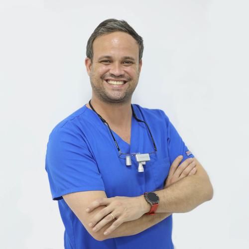
Oral surgeon, implantologist
17 years in practice
CBCT, OPG or periapical: which one you actually need
| Examination |
What it answers |
When it is the right one |
| Periapical X-ray |
One or two teeth in close detail, flat |
Decay, checking a root filling, pain localised to a single tooth |
| OPG (panoramic) |
Both arches at a glance, also flat |
General review, counting teeth, a first look at the wisdom teeth |
| Dental CBCT |
A 3D volume with real measurements |
Implants, surgery, wisdom teeth near the nerve, difficult root canals, orthodontics with buried teeth |
The usual order runs from least to most: start with whatever carries the lowest dose and only step up when it fails to answer the question. Going straight to the 3D study makes sense when the indication is already clear — implant planning, say — not as the opening move of a routine check-up.
Who we image
Our own patientsWhere it is part of the diagnosis and the treatment plan
Referrals from other dentistsWe do the imaging, you go back to them with the file
Second opinionsAnyone wanting to check an implant plan proposed elsewhere
Visitors and short staysImaging and interpretation the same day, in one trip
Previously operated casesFollowing up grafts, sinus lifts and implants placed years ago
Four languagesEnglish, Spanish, Russian and Ukrainian, with no interpreter needed
Where we are and how to get here
The imaging room is inside the clinic, at c/ Entença, 69, in the Esquerra de l'Eixample, two minutes from Rocafort station on metro line L1. Images are taken in the same place the dentist sees you, so there is no appointment to book elsewhere and nothing to come back for another day.
| Address |
c/ Entença, 69 · 08015 Barcelona |
| Nearest metro |
Rocafort, line L1 |
| Scanning hours |
Monday to Thursday 10:00–19:00, Friday 9:00–18:00 |
Getting to us
We are where Entença crosses the lower part of the left Eixample, between Gran Via and Avinguda de Roma. The simplest landmark is line L1: Rocafort is our stop, and Urgell and Espanya either side of it are both walkable. Coming in on L3, get off at Tarragona; on L5, at Entença, which is the top end of our own street; from Sant Antoni it is a short walk from the L2 station of that name. Driving, the car park is at Entença 61, same block. In a taxi, Entença with Gran Via is enough to say.
Where people travel in from
An imaging appointment is usually a one-off and often a long way from home, sometimes with another dentist's referral in hand. The clinic sits on the western edge of the district, next to the Sants interchange and Plaça d'Espanya, which makes the journey shorter from the south and from the Baix Llobregat than going into the centre would be.
| Coming from |
Route |
| Neighbouring districts |
Sant Antoni, Poble-sec, Hostafrancs, Sants and both halves of the Eixample: on foot or a couple of metro stops |
| L'Hospitalet, Santa Coloma |
L1 runs straight to Rocafort, no changes |
| Cornellà, Esplugues, Sant Joan Despí |
L5 as far as Entença station, then down the street |
| Sant Boi, Sant Vicenç dels Horts, Pallejà |
FGC on the Llobregat-Anoia line to Espanya, the station next to ours |
| Martorell, Abrera, Olesa, Manresa, Igualada |
The same FGC line to Espanya, then one metro stop |
| El Prat, Castelldefels, Gavà, Viladecans, Sitges, Vilanova |
Rodalies train to Sants and change onto L1 |
| Badalona, El Masnou, Premià, Mataró |
The Maresme line into Sants and one change |
| Terrassa, Sabadell, Granollers, Mollet |
Any suburban line to Sants or Plaça Catalunya, then L1 |
| Girona, Tarragona, Lleida |
Long-distance trains arrive at Sants, one metro stop away |
Travelling in from outside the city, or over for a few days? Mention it when you book: imaging and the discussion go back to back, so you leave with the images and a plan rather than making the trip twice.
The clinical team
The images are read by the dentist who will be handling the case. Depending on what it concerns — implants, root canals, orthodontics or children's dentistry — they are reviewed by whoever works in that area.
Why have it done here
One appointmentIndication, imaging, interpretation and plan in a single sitting rather than spread across three addresses.
The file leaves with youIn DICOM and with a viewer, whether or not you are treated here.
Smallest useful fieldWe cover what answers the question, not both arches as a matter of routine.
The machine is namedCarestream CS 8200 3D Advance Edition, inside the clinic rather than subcontracted out.
Price fixed beforehandThe figure is quoted before anything is switched on, and no VAT is added to it.
English spoken throughoutReception, the appointment and the explanation of the findings.
Cases
Section in preparation.
Common questions
Can I just have the scan without being treated at the clinic?
Yes. It commits you to nothing: you pay, we do it, you get the file. The only thing that cannot be skipped is a written indication, yours or your dentist's, because radiation protection rules do not allow X-rays without one.
Do I need a referral from my dentist?
If you have one, bring it and we cover the field it asks for. If you do not, the indication is established at a first appointment, which costs €50, and the scan follows the same day where it is warranted.
Is this the same as what the hospital does?
No. What a dental clinic has is a CBCT: jaws only, sitting up, seconds, no contrast, and a far lower dose. A hospital CT covers any part of the body with a different beam geometry and a different dose, and for implants or orthodontics it is not the right examination.
Will my dentist at home be able to open the file?
Yes. It comes as DICOM, which is the international standard every dental planning package reads, so it loads wherever you take it. If your dentist wants a specific export instead, let us know when you book.
Does it hurt or feel claustrophobic?
Neither. Your chin rests on a support and you hold still for one turn of the arm. Nothing goes in your mouth, nothing encloses you, and there is no noise beyond the motor.
How long until I get the results?
The reconstruction finishes in under a minute and is gone through with you in the same appointment. The file is handed over the same day, as a download link or on a USB stick.
I had a dental CT scan two years ago — will it do?
Not for planning surgery. Past the one-year mark the bone and the position of the teeth have changed enough that the measurements stop being reliable. Bring it anyway: next to a new one it shows how things have moved.
Can it be done during pregnancy?
It waits, unless something urgent cannot. If you are pregnant or suspect you might be, say so before going into the room — the decision is made case by case, and anything urgent is discussed with your obstetrician.
And for children?
Only where it will alter what gets done, and always at the narrowest field. Most of what comes up in children's dentistry is settled with an ordinary X-ray, which carries less dose.
Do I need to fast beforehand?
No. Nothing is injected, so eat and drink as you normally would.
Book a dental CT scan
Message us on WhatsApp and we will tell you which examination your case needs and what it costs before you come in.
Message us on WhatsApp c/ Entença, 69 · Barcelona · Monday to Thursday 10:00–19:00, Friday 9:00–18:00
This page is for information and does not replace an appointment. Any X-ray examination is indicated by a dentist or doctor after examining the patient. Prices current for 2026.